


Modern operations to restore vision are high-tech and safe procedures that can eliminate almost any ophthalmological problem.They have been successfully used for several decades, so the methods are constantly developing, expanding and becoming more effective.Improving visual functions is achieved using hardware correction of the shape of the cornea, lens, retina and other elements of the optical system of the eye.Properly selected technology allows not only to completely restore vision, but also to reduce the risk of complications.From the article you will learn what ophthalmological operations exist, indications for use and possible risks.
Species
Thanks to the development of hardware methods of medicine, operations to restore vision today are reliable and minimally invasive procedures.Their duration does not exceed several hours, and in the future there is no need for complex rehabilitation measures.The choice of surgical treatment method is chosen depending on the disease, age and general condition of the patient’s visual system.
Laser correction
The most popular type of surgery to correct visual acuity.Today these are sophisticated high-tech methods that are highly effective and have minimal risk of complications.Allows you to cope with myopia, farsightedness and astigmatism.After the procedure, visual acuity is maintained for a long time, and if you follow all the ophthalmologist’s instructions, you can completely avoid repeated intervention.There are several types of laser correction:
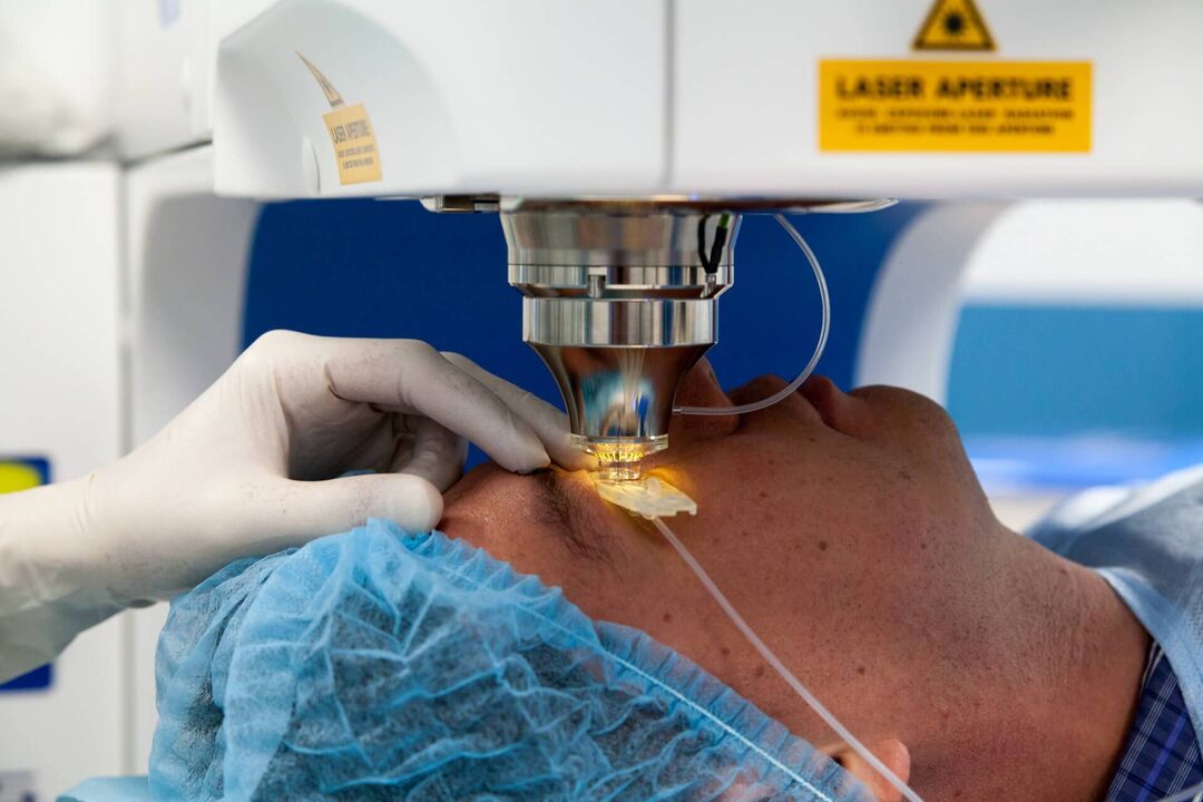
- LASIK.Basic type of surgery to restore visual acuity.First, the surface layer of the cornea is separated with a microkerat, and then its shape is changed using a laser beam.The main disadvantage of this type of correction is the inability to take into account the individual characteristics of the patient’s eye anatomy;
- Super LASIK.An improved version of the traditional LASIK technique.Allows you to achieve a better result, since it takes into account the structure of the patient’s visual system.Used in most modern clinics in the world;
- Femto LASIK.A similar type of operation, the only difference is that the cornea is cut not with a microkerat, but with a special femto laser.There is also an improved version, in which the course of the operation depends on the individual characteristics of the patient - Super Femto LASIK;
- Epi-LASIK.The mechanism of the procedure is identical to the traditional LASIK method, but this operation is prescribed only to patients with a thinned cornea (acquired or congenital);
- PRK (FRK).Photorefractive keratectomy has been performed since 1985.Today it is used when there are contraindications to conventional correction methods, for example, with a thin cornea or serious ophthalmological diseases.The healing process is always painful, and the recovery period lasts longer than with other methods.
Vision correction operations last no more than 15 minutes.After the procedure, it is necessary to wear a protective bandage for several hours, as well as instill drops for 1-2 months.The risk of complications is minimal; re-treatment is necessary if there is a significant decrease in vision.
Vitrectomy
This is a procedure for complete or partial removal of the vitreous humor of the eyeball.It is performed under general or local anesthesia; in the absence of complications, it resolves in 2-3 hours.First, small punctures are made in the eye socket, through which subsequent manipulations are carried out.As a rule, this involves laser cauterization of the affected areas of the retina, compaction of detachments, or restoration of tissue integrity.The procedure is prescribed for the following problems:
- restoration of visual functions after hemorrhage in the tissues of the eye;
- prevention of age-related retinal detachment;
- Treatment of severe eye retinopathy that causes rough scarring or neovascularization (growth of blood vessels).
Artificial polymers, a gas bubble, silicone oil, or a balanced salt solution are used as vitreous replacements.The latter type is used more often, since further surgery is not required - the saline solution is subsequently replaced by intraocular fluid.
After surgery, side effects are possible in the form of corneal edema, increased intraocular pressure, or even further decreased vision.Recovery and prognosis depend on the extent of the lesion, as well as the type of prosthesis used to replace the vitreous.If there are irreversible changes in the optic nerve, then vision correction is almost impossible to achieve.
Scleroplasty
A common ophthalmological procedure aimed at strengthening the outer layer of the eye (sclera).It is prescribed not to correct visual functions, but to stabilize the degree of myopia in a patient at risk.It is recommended for teenagers suffering from this problem, since at this age the shape of the eye actively changes.
During the operation, the required number of flaps of material are inserted behind the back wall of the eyeball to strengthen the sclera.Typically polymers or biological components are used.After this, adhesion occurs to the outer shell of the eye, and after a few months, blood vessels necessary to maintain visual functions grow into the flap.There is also a simplified version of scleroplasty.It involves the introduction of an artificial or biological substance behind the eyeball.The mechanism of action of this technology is identical - preventing the growth of the eyeball.
This is a well-studied operation that has remained virtually unchanged over the years.It is carried out in most clinics.There are practically no side effects identified, with the exception of a possible allergy to the drug.Repeat surgery is usually required.
Lens replacement
A necessary operation that is prescribed for clouding or any other degenerative processes in the lens, for example, cataracts.Treatment is always forced, but the implant is selected individually, depending on age, gender and the severity of pathological changes in the eye.Lens replacement is prescribed in the following cases:
- high degrees of myopia and farsightedness;
- significant decrease in refraction;
- regenerative processes in the eye, age-related vision loss;
- impossibility of laser vision restoration;
- cataract;
- the likelihood of developing glaucoma against the background of a systemic or ophthalmological disease.
The procedure is always performed under local anesthesia.During the operation, the surgeon makes a small incision with a laser, after which a special tool liquefies the patient's lens and removes it from the eye.After this, the prepared graft is installed.The intervention lasts no more than 25 minutes; subsequent suturing and recovery in a hospital setting are not required.
The operation is performed in most private and public clinics.Complications after manipulation are usually not observed, but subsequent laser vision correction is often prescribed.In rare cases, the lens needs to be replaced again.
Keratoplasty (cornea replacement)
One of the most modern and complex ophthalmological operations, which is associated with many risks and requires a highly qualified surgeon.Required to restore the anatomical integrity and physiological functions of the cornea.Prescribed for the treatment of congenital or acquired defects resulting from injury or disease.Healthy tissue for transplantation is taken only from donors, but the development of artificial replacement is underway in many countries.Keratoplasty is recommended to solve the following problems:
- treatment of corneal diseases (sores, tone disorders);
- mechanical or chemical damage;
- birth defects.
The operation takes no more than 30 minutes.During the procedure, the surgeon uses a laser or a special scalpel to remove part of the patient’s cornea and implant donor tissue in its place.The stitches can last up to a year, after which a special lens is selected to reduce the risk of infection.The recovery period is from 4 weeks, during which antibiotic instillations are necessary, but regular examinations are required throughout the next year.
In recent years, it has been possible to significantly reduce the risk of donor tissue rejection due to the use of special compounds during its processing and preservation.
Laser coagulation of the retina
Surgical method for retinal tissue restoration.The effectiveness of the method is more than 70%, and within 24 hours after its implementation you can return to your normal lifestyle.Observations by an ophthalmologist are required for a year after the procedure.
Today, the operation is performed using a laser, which eliminates the need for blood loss.It is carried out under local anesthesia, the procedure takes no more than 20 minutes.
Before exposure to the laser, drops are instilled to dilate the pupil, and then a special protective lens is put on, through which the exposure occurs at low frequencies.Due to high temperatures, damaged cells and small blood vessels stick together.
The coagulation procedure is necessary for any damage and pathologies of the retina, as well as for eye tumors and diseases of the vascular system of this organ.After surgery, inflammation and cloudiness may develop.For several years after correction, you should not engage in heavy physical labor or active sports.
Crosslinking
An effective method for treating various corneal diseases.It is carried out to strengthen ligaments and other fibers in the corneal tissue, which is necessary for keratoconus of varying degrees or degenerative processes, dystrophy.
The operation is performed under local anesthesia.First, a small part of the cornea is cut off with a special device, and vitamin B2 is instilled into the open area.Subsequent irradiation makes it possible to tighten the tissue by more than 200%.You must wear a protective contact lens for the first week after surgery, and be examined by a doctor for 6 months.The effect of the procedure lasts 10 years, then a second operation is required.
Complications are observed in rare cases.The patient may experience decreased vision, inflammation or clouding of the cornea.
Glaucoma treatment
Ophthalmic surgery for various degrees of glaucoma is necessary when drug therapy does not bring the desired result.The operation is performed using a laser or surgically.
The laser method is considered the most successful.It is completely painless for the patient, and there are practically no complications.During the procedure, a hole is made with a beam through which fluid is removed from the tissues of the eye to normalize pressure.Used to treat all types of glaucoma.
Manual surgery is less effective because there is a risk of complications after the procedure.Typically, this is a non-penetrating deep sclerectomy.The purpose of the method is to reduce intraocular pressure by slightly thinning the corneal layer.
The effect after both types of operations decreases over time.On average, repeat surgery is required after 5-7 years.This period can be extended with the help of competent drug treatment.
Conclusions
Today, most modern ophthalmology clinics perform a number of surgical procedures to correct vision.These are precise, high-tech methods that can be used to eliminate almost any eye defect.The choice of method depends on a number of factors – age, disease, individual structural features of the patient’s visual system.After the operation, the effect occurs almost immediately, and if all the doctor’s instructions are followed, visual acuity can be maintained for a long time.